After a mammogram, some women find out they have dense breasts. They come to me with questions about what that means, how it affects their risk for breast cancer, and what they should do differently. Here’s how I answer the questions I hear most often.
What does it mean to have dense breasts?
It’s common for women to have dense breasts. Your breasts are made of fatty tissue, which is not dense, and supportive tissue, milk glands, and milk ducts, which is. The parts of your breast made up of dense tissue show up as white on a mammogram, so it can be harder to spot signs of breast cancer in those areas.
How do I know if I have dense breasts?
The radiologist who reviews your mammogram assigns a grade to your breast density based on how much of your breast tissue is dense. You might see something on your mammogram report called Breast Imaging Reporting and Data System (BI-RADS). There are four levels of breast density:
- A is almost all fatty tissue, found in about 10 percent of women
- B is more nondense than dense, found in about 40 percent of women
- C is more dense than nondense, found in about 40 percent of women
- D is almost all dense, found in about 10 percent of women
If you fall into the C or D categories, your mammogram report may indicate that you have dense breasts. If it doesn’t say, ask your doctor.
As you can see, about half of all women have dense breasts. You’re more likely to have dense breasts if you are younger, have less body fat, and/or take hormone therapy for menopause.
How does my breast density affect my risk for breast cancer?
Since it’s harder to spot breast cancer on dense breasts, you have a higher chance of cancer not being detected on a mammogram. Separately from that, women with dense breasts also have a higher risk of breast cancer.
What should I do differently if I have dense breasts?
You should talk to your doctor about your other risk factors for breast cancer and work together to come up with a breast cancer screening schedule that works for you. For my patients with dense breasts but no additional risk for breast cancer, I recommend an annual mammogram beginning at age 40. Depending on other risk factors for breast cancer, I might also recommend:
- A breast MRI, which uses magnetic forces to image your breast
- A 3D mammogram, which combines images of your breast taken from different angles
- Breast ultrasound, which uses sound waves to investigate areas of your breast that might be concerning
- Molecular breast imaging, which uses a radioactive tracer to look for cancerous areas

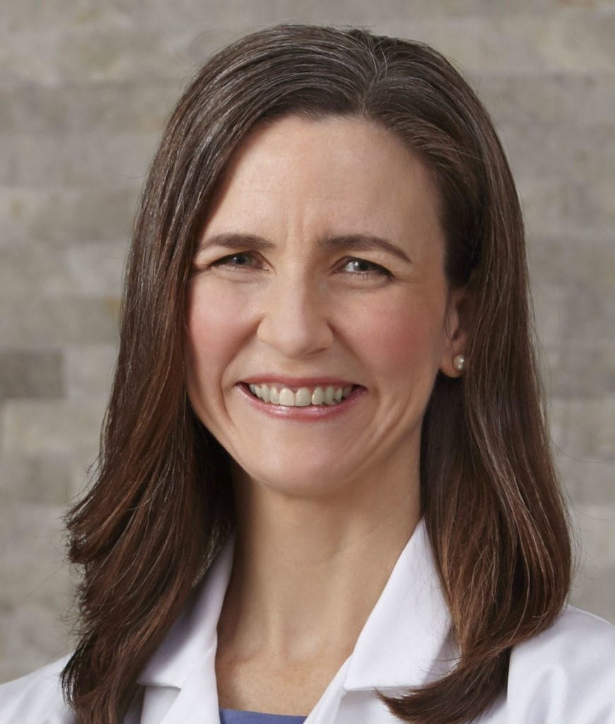
Valerie Gorman, MD, FACS, is a breast cancer surgeon. She is board certified by the American Board of Surgery and serves as Chief of Surgery and Medical Director of Surgical Services at Baylor Scott & White Medical Center – Waxahachie. She is the Clinical Assistant Professor of Medical Education position at the Texas A&M University College of Medicine.
- Certificate, Physician Leadership Program, Southern Methodist University, Dallas, Texas (2010)
- M.D., University of Texas Southwestern Medical School at Dallas, Texas (June 1999)
- B.S., Biola University, LaMirada, California, (1994) Magna Cum Laude
Major: Biochemistry - Residency in General Surgery, University of Texas Southwestern Medical Center at Dallas, Texas (June 2004)